Microplate Sonication in Shotgun Proteomics – Application Note
Shotgun proteomics depends on efficient, reproducible sample preparation to convert complex biological material into LC-MS/MS-ready peptides. The UIP400MTP microplate sonicator supports this process by enabling standardized, parallel sonication of small-volume samples, helping laboratories improve disruption, extraction, and resolubilization in microplate-based workflows. This application note describes how the UIP400MTP can be integrated into proteomic sample preparation, using a published extracellular-vesicle workflow from PLA2G12A/Th17 studies as a lab-tested example.
Microplate Sonication for More Reproducible Shotgun Proteomics
For proteomics researchers, reproducible sample preparation is critical for high-quality LC-MS/MS data. The UIP400MTP microplate sonicator supports standardized, parallel sonication in plate-based and small-volume/high-throughput workflows.
It is especially useful for laboratories working with:
- Large sample sets that require consistent processing across many wells
- Limited-input material, where sample loss and variability must be minimized
- Lipid-rich samples, such as extracellular vesicles
- Complex biological preparations that require efficient disruption, extraction, or resolubilization
- Comparative shotgun proteomics, where reproducibility across conditions and replicates is essential
By integrating microplate sonication before digestion, researchers can improve sample readiness for:
- Protein extraction and resolubilization
- Trypsin/Lys-C digestion
- Nano-LC/MS/MS acquisition
- Quantitative downstream proteomics analysis
Learn how microplate sonication helps reduce manual variability, improve sample disruption, and prepare complex biological samples for reliable LC-MS/MS analysis.

Microplate sonication can benefit:
- Proteomics core facilities
- EV research laboratories
- Immunology and cell-biology labs
- Translational research groups
- Life-science teams scaling from single-sample prep to higher-throughput workflows
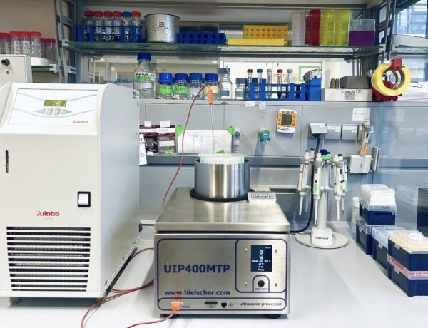
The UIP400MTP is not an ultrasonic bath. It'a a high intensity cup horn for focused sonication. This powerful non-contact sonicator delivers a uniform sonication across all wells of your standard well-plate. You have precise control over amplitude, power, and pulsing. A built-in timer and temperature probe ensures consistent results. The UIP400MTP plate sonicator cools samples with water bath (external chiller optional).
The UIP400MTP is not an ultrasonic bath. It'a a high intensity cup horn for focused sonication. This powerful non-contact sonicator delivers a uniform sonication across all wells of your standard well-plate. You have precise control over amplitude, power, and pulsing. A built-in timer and temperature probe ensures consistent results. The UIP400MTP plate sonicator cools samples with water bath (external chiller optional).
High-Throughput Sample Preparation for Extracellular Vesicle Proteome Analysis
What is Shotgun Proteomics?
Shotgun proteomics has become a central analytical strategy for characterizing complex biological samples, from whole-cell lysates and tissue extracts to purified organelles, extracellular vesicles, and low-input clinical specimens. Its core strength lies in the coupling of protein digestion, high-resolution liquid chromatography-tandem mass spectrometry, and computational peptide-to-protein inference. However, the quality of the final proteome dataset is determined long before the sample reaches the mass spectrometer. Efficient sample disruption, protein solubilization, removal of interfering substances, reproducible enzymatic digestion, and robust peptide recovery are all decisive for depth of coverage and quantitative reliability.
For proteomics researchers and life-science laboratories working with limited or precious samples, sample preparation is often the bottleneck.

The UIP400MTP ensures reliable sample preparation and a facile integration with existing lab workflows
The Challenge of Extracellular Vesicles in Proteomics
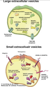 Extracellular vesicles (EVs), for example, represent a particularly demanding sample class. They are membrane-bound, lipid-rich, nanoscale particles with relatively low protein yield and a high potential for carry-over of lipids, salts, detergents, serum proteins, and other matrix components. These features can interfere with digestion efficiency, chromatographic performance, electrospray stability, and peptide identification. A preparation workflow for EV proteomics must therefore be strong enough to disrupt vesicle structures and solubilize protein cargo, while remaining compatible with downstream enzymatic digestion and LC-MS/MS analysis.
In this context, microplate-compatible ultrasonication offers a practical approach for reproducible, parallelized sample processing. The Hielscher microplate sonicator models UIP400MTP (400 W) and UIP550MTP (550 W) are designed for sonication of samples in multiwell plates and small-volume vessels, supporting higher-throughput workflows than conventional single-probe sonicators. For proteomics laboratories, this format is attractive because it can reduce hands-on variability, improve parallel treatment of multiple samples, and integrate more naturally into plate-based sample preparation pipelines.
Exemplary Workflow
A recent extracellular vesicle proteomics workflow in PLA2G12A-driven Th17-cell biology provides a useful, lab-tested example of how the microplate sonicator UIP400MTP can be incorporated into shotgun proteomics sample preparation. In that study series, EV samples were processed for proteome analysis using methanol treatment, UIP400MTP sonication, centrifugation, drying, trypsin/Lys-C digestion, and nano-flow LC-high-resolution tandem MS. The biological work was reported in 2026 by Mochizuki-Ono and colleagues, where the proteomics analysis helped characterize how PLA2G12A alters EV cargo proteins in the context of pathogenic T-cell responses.

96-well plate sonicator UIP400MTP for high-throughput protein extraction
Sonicator: UIP400MTP microplate sonicator
Input: 5 µg EV protein equivalent
Digestion: Trypsin/Lys-C
Readout: Nano-LC-MS/MS / DIA proteomics
Step-by-Step Instruction: UIP400MTP-Assisted EV Sample Preparation for Shotgun Proteomics
This workflow describes a low-input extracellular vesicle (EV) shotgun proteomics sample preparation method using the UIP400MTP microplate sonicator. It is based on a published EV proteomics workflow in which EV samples were normalized, dried, treated with methanol, sonicated, digested with trypsin/Lys-C, acidified, and analyzed by nano-LC-MS/MS.
The procedure is intended for proteomics researchers and life-science laboratories preparing EVs or other small-volume, lipid-rich biological samples for bottom-up shotgun proteomics.
Workflow Overview
| Stage | Purpose | Key Output |
|---|---|---|
| EV preparation | Isolate, wash, and quantify EV samples | Normalized EV input |
| Sample drying | Remove liquid before solvent-assisted processing | Dried EV material |
| Methanol treatment | Support disruption of lipid-rich EV material | Methanol-wetted sample |
| UIP400MTP sonication | Promote disruption, extraction, and homogenization | Processed EV sample |
| Digestion | Generate peptides from EV proteins | Trypsin/Lys-C peptide digest |
| LC-MS/MS analysis | Separate, detect, and quantify peptides | Proteomics dataset |
| Data analysis | Identify and quantify peptides and proteins | Protein-level results |
Step-by-Step Protocol
| Step | Instruction | Critical Parameters |
|---|---|---|
| 1 | Prepare purified EV samples using the laboratory’s established isolation workflow. | Remove cells, debris, and major contaminants before proteomics preparation. |
| 2 | Quantify EV protein content, for example by BCA assay. | Normalize all samples to equal protein-equivalent input. |
| 3 | Transfer 5 µg EV protein equivalent into clean PCR tubes or compatible low-volume tubes. | Use low-bind tubes where sample loss is a concern. |
| 4 | Dry the EV samples completely. | Avoid overheating; confirm that no visible liquid remains. |
| 5 | Add 25 µL methanol to each dried EV sample. | Ensure the dried material is fully wetted. |
| 6 | Sonicate the samples for 3 minutes using the UIP400MTP microplate sonicator. | Use identical sonication settings and layout logic across all samples. |
| 7 | Centrifuge the sonicated samples at 19,000 × g for 20 minutes at 4°C. | Avoid disturbing any pellet or insoluble material after centrifugation. |
| 8 | Carefully remove 20 µL of the supernatant. | Use consistent pipetting technique across all samples. |
| 9 | Dry the remaining sample material completely. | Proceed directly to digestion or store only under validated conditions. |
| 10 | Add 4 µL trypsin/Lys-C solution at 100 ng/µL in 50 mM ammonium bicarbonate. | Prepare enzyme solution fresh or use properly stored aliquots. |
| 11 | Sonicate briefly to dissolve the dried protein material in digestion solution. | Collect all liquid at the bottom of the tube after sonication. |
| 12 | Digest for 2 hours at 37°C with shaking at 300 rpm. | Keep caps tightly closed to prevent evaporation. |
| 13 | Add 1 µL of 1.25% TFA to acidify the digest. | Acidification stops digestion and prepares peptides for LC-MS/MS. |
| 14 | Transfer the digest to LC-MS-compatible vials or plates. | Avoid transferring insoluble debris. |
| 15 | Analyze by nano-LC-MS/MS. | Use consistent injection volume, LC gradient, and MS acquisition settings. |
| 16 | Process raw data using DIA, DDA, or targeted proteomics software as appropriate. | Apply suitable database, enzyme specificity, modification settings, and FDR thresholds. |
(cf. Mochizuki-Ono et al., 2026)
Multi-well plate sonicator UIP400MTP
Why microplate sonication?
The UIP400MTP enables standardized, parallel sonication of small-volume samples. This is useful when reproducibility, low sample input, and multi-sample throughput are important.

Use any standard microplate! High-throughput sample prep with the UIP400MTP
The referenced EV proteomics workflow used 5 µg protein-equivalent EV input, 25 µL methanol, 3 minutes of UIP400MTP sonication, trypsin/Lys-C digestion, TFA acidification, and nano-LC-MS/MS analysis.
Recommended LC-MS/MS and Data Analysis Steps
After digestion and acidification, samples can be analyzed using nano-flow reversed-phase LC coupled to high-resolution tandem mass spectrometry. In the published workflow, peptides were separated on a nano-LC system and analyzed by data-independent acquisition on an Orbitrap mass spectrometer.
| Stage | Recommendation | Purpose |
|---|---|---|
| Sample loading | Use a C18 trap or equivalent peptide-loading setup. | Concentrates peptides and removes highly polar contaminants. |
| Peptide separation | Use nano-flow reversed-phase LC. | Improves peptide separation and MS sensitivity. |
| MS acquisition | Use DIA, DDA, or targeted MS depending on study design. | Generates peptide-level spectral data. |
| Database search | Use a species-appropriate reference database. | Supports peptide and protein identification. |
| FDR control | Apply peptide and protein false-discovery-rate thresholds. | Controls identification confidence. |
| Quantification | Export peptide, precursor, and protein abundance tables. | Enables statistical comparison between groups. |
Quality Control Checklist
- Confirm equal EV protein-equivalent input across all samples.
- Use randomized or balanced sample processing layouts.
- Keep methanol volume, sonication time, drying time, and digestion volume consistent.
- Monitor peptide yield and total protein identifications.
- Check missed-cleavage rate and peptide length distribution.
- Inspect chromatographic peak shape and retention-time reproducibility.
- Include blanks to monitor carryover.
- Use pooled QC samples for larger studies.
- Evaluate replicate coefficient of variation.
- Use PCA or related methods to identify batch effects or outlier samples.
Protocolling Recommendations
 When publishing or documenting this workflow, report the EV input amount, plate format, methanol volume, amplitude and sonication time of UIP400MTP, centrifugation conditions, drying method, digestion buffer, enzyme concentration, digestion time, acidification conditions, LC-MS/MS platform, acquisition mode, software version, database, FDR threshold, normalization method, and statistical workflow.
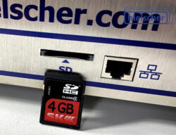
All Hielscher microplate sonicators automatically record important process parameters such as amplitude, sonication duration and temperature with date and time stamp as a CVS file on the integrated SD-card. Data protocoling for reproducibility and quality control was never easier!
For DIA proteomics, use consistent acquisition settings across all samples and include pooled QC samples where possible. Monitor precursor counts, retention-time stability, and replicate variability.
This workflow is a lab-tested example for EV proteomics. For other sample types, optimize input amount, solvent conditions, sonication time, digestion volume, and LC-MS/MS injection strategy before scaling to a full study.
The Study in Detail: EV Shotgun Proteomics in PLA2G12A Study
The PLA2G12A study provides a concrete example of UIP400MTP use in a shotgun proteomics workflow. The biological question was how the secreted phospholipase PLA2G12A modifies Th17-cell-derived extracellular vesicles and thereby influences pathogenic T-cell differentiation. The authors showed that PLA2G12A acts on EV membranes to produce lysophospholipids, including 1-oleoyl-lysophosphatidylethanolamine, and that downstream lysophosphatidic acid signaling via LPA2 contributes to Th17 differentiation. Beyond lipid signaling, the studies also examined EV cargo, including RNA and protein content, making shotgun proteomics an important component of the characterization strategy.
In the EV preparation workflow, Th17 cells were cultured in medium containing exosome-depleted fetal bovine serum. Culture supernatants were first centrifuged to remove cells, filtered through a 0.22 µm filter, concentrated by ultrafiltration/ultracentrifugation, washed, resuspended in PBS, and quantified by BCA protein assay. This upstream EV preparation ensured that a defined protein-equivalent amount of EV material could be carried into the proteomics workflow.
For shotgun proteomic analysis, the authors used EV samples corresponding to 5 µg protein equivalents. These samples were dried in PCR tubes, and 25 µL methanol was added. The samples were then sonicated for 3 minutes using the UIP400MTP microplate sonicator. Following sonication, the samples were centrifuged at 4°C for 20 minutes at 19,000 × g. After removal of 20 µL supernatant, the samples were centrifuged to dryness. Proteins were then dissolved by sonication in 4 µL trypsin/Lys-C solution at 100 ng/µL in 50 mM ammonium bicarbonate. Digestion was performed for 2 hours at 37°C with shaking at 300 rpm. The digest was acidified with 1 µL of 1.25% trifluoroacetic acid and submitted to nano-flow LC-high-resolution tandem mass spectrometry.
This workflow highlights several useful principles for low-input EV proteomics. First, the input was normalized by protein equivalent, which is important when comparing EVs from different genotypes or treatments. Second, methanol was used before sonication, supporting disruption and extraction while avoiding detergent systems that may complicate LC-MS/MS. Third, the sonication step was short and standardized, which is compatible with parallel sample processing. Fourth, trypsin/Lys-C digestion was performed in a very small volume, reducing dilution and supporting low-input peptide recovery. Finally, direct transition from digestion to acidification and LC-MS/MS minimized unnecessary handling steps.
The downstream LC-MS/MS method used nano-flow reversed-phase separation coupled to a Q-Exactive HF Orbitrap mass spectrometer. Samples were trapped on a C18 pre-column, then separated on a 50 µm inner-diameter analytical column at 200 nL/min and 40°C. The gradient ran from low organic solvent to 35% solvent B over 60 minutes, followed by high-organic washing and re-equilibration. MS acquisition was performed in positive-ion mode using data-independent acquisition with higher-energy collisional dissociation. DIA raw files were analyzed using DIA-NN 1.8 with an in silico predicted spectral library.

High-throughput protein extraction with the 96-well plate sonicator UIP400MTP
Frequently Asked Questions
Who benefits most from microplate sonication in shotgun proteomics?
Microplate sonication is especially useful for proteomics laboratories processing multiple samples in parallel, including core facilities, proteome research groups, immunology labs, translational research teams, and life-science laboratories moving toward higher-throughput LC-MS/MS workflows. It is particularly relevant when sample-to-sample reproducibility is critical.
Why use the UIP400MTP instead of a conventional probe sonicator?
A conventional probe sonicator typically processes samples one at a time and requires careful cleaning between samples to reduce carryover. The microplate sonicator models UIP400MTP and UIP550MTP support parallel sonication in a plate-based or small-volume format, helping reduce manual handling, improve consistency, and streamline multi-sample preparation. They allow for easy integration into automated workflows, too.
Where does sonication fit into the shotgun proteomics workflow?
Sonication is usually applied during the pre-digestion sample-preparation phase. It can support disruption, extraction, homogenization, and resolubilization before enzymatic digestion with trypsin, Lys-C, or a trypsin/Lys-C mixture.
Additionally, sonication can be also applied during digestion to speed up enzymatic protein digestion significantly. Discover the potential of high-throughput protein digestion using sonication for a fast proteomic workflow!
Is microplate sonication useful for extracellular vesicle proteomics?
Yes. Extracellular vesicles are lipid-rich, membrane-bound particles that can be challenging to process reproducibly. In the referenced PLA2G12A studies, EV samples were treated with methanol, sonicated with the UIP400MTP, dried, digested with trypsin/Lys-C, and analyzed by nano-LC/MS/MS.
What sample types can benefit from microplate sonication?
Microplate sonication can be useful for cell lysates, organelle fractions, immunoprecipitates, protein aggregates, membrane-rich samples, extracellular vesicles, and other low-volume biological preparations. Each sample type should be optimized for sonication intensity and time, solvent conditions, input amount, and digestion strategy.
Does sonication replace enzymatic digestion?
No. Sonication helps prepare the sample for digestion by improving disruption, extraction, or resolubilization. Proteolytic digestion is still required to generate peptides for bottom-up shotgun proteomics.
Read more about ultrasonically promoted protein digestion in proteomic workflows!
Is the UIP400MTP compatible with low-input proteomics?
Yes, the microplate format is well suited to small-volume workflows where sample conservation and consistent processing are important. In the published EV workflow, the proteomics preparation was performed with 5 µg protein-equivalent EV input.
Can microplate sonication improve reproducibility?
Hielscher sonicators support reproducibility by applying standardized sonication conditions across multiple samples. However, other factors such as cell or EV isolation, input normalization, digestion efficiency, LC-MS/MS stability, data processing, and statistical analysis contribute to overall proteomics reproducibility, too.
Does microplate sonication improve protein identification depth?
It may contribute to improved identification depth when sample disruption or resolubilization is a limiting factor. The effect depends on sample type, protein chemistry, cleanup strategy, digestion conditions, and LC-MS/MS method performance.
What should be optimized before using the workflow routinely?
Key parameters include sample input, buffer or solvent composition, plate format, ultrasonic amplitude and duration, drying conditions, digestion volume, enzyme concentration, incubation time, and LC-MS/MS injection amount. Pilot testing is recommended before applying the workflow to a full experimental set.
Can this workflow be used with DIA proteomics?
Yes. The referenced EV workflow used data-independent acquisition followed by DIA-NN analysis. Microplate sonication is part of the upstream sample-preparation process and can be integrated with DIA, DDA, or targeted proteomics methods.
What quality-control metrics should be monitored?
Recommended QC metrics include peptide yield, number of identified peptides and protein groups, missed-cleavage rate, peptide length distribution, chromatographic peak shape, retention-time stability, replicate coefficient of variation, carry-over, and separation of biological groups by PCA or related methods.
What is the main advantage of integrating the microplate sonicator models UIP400MTP or UIP550MTP into proteomics sample preparation?
The main advantage is standardized, parallel processing of small-volume samples. This helps laboratories reduce manual variability and prepare complex biological samples more consistently for digestion, LC-MS/MS acquisition, and quantitative proteomics analysis.
Hielscher microplate sonicators can be seamlessly integrated into automated workflows.
Literature / References
- FactSheet UIP400MTP Multi-well Plate Sonicator – Non-Contact Sonicator – Hielscher Ultrasonics
- FactSheet UIP550MTP Multi-well Plate Sonicator – Non-Contact Sonicator – Hielscher Ultrasonics
- Mochizuki-Ono C., Taketomi Y., Irie A., Kano K. et al. (2026): PLA2G12A-driven extracellular vesicle-lipid signaling amplifies pathogenic T cell responses in inflammatory diseases. Cell Reports 45, 2026.
- Mochizuki, Chika; Taketomi, Yoshitaka; Irie, Atsushi; Kano, Kuniyuki; Nagasaki, Yuki; Miki, Yoshimi; Ono, Takashi; Nishito, Yasumasa; Nakajima, Takahiro; Tomabechi, Yuri; Hanada, Kazuharu; Shirouzu, Mikako; Watanabe, Takashi; Hata, Kousuke; Izumi, Yoshihiro; Bamba, Takeshi; Chun, Jerold; Kudo, Kai; Kotani, Ai; Murakami, Makoto (2024): Secreted phospholipase PLA2G12A-driven lysophospholipid signaling via lipolytic modification of extracellular vesicles facilitates pathogenic Th17 differentiation. BioRxiv 2024.
- Lischnig A., Bergqvist M., Ochiya T., Lässer C. (2023): Corrigendum for “Quantitative Proteomics Identifies Proteins Enriched in Large and Small Extracellular Vesicles”. Molecular & Cellular Proteomics, 22; 2023.
- Lauren E. Cruchley-Fuge, Martin R. Jones, Ossama Edbali, Gavin R. Lloyd, Ralf J. M. Weber, Andrew D. Southam, Mark R. Viant (2024): Automated extraction of adherent cell lines from 24-well and 96-well plates for multi-omics analysis using the Hielscher UIP400MTP sonicator and Beckman Coulter i7 liquid handling workstation. Metabomeeting 2024, University of Liverpool, 26-28th November 2024.
- Cosenza-Contreras M, Seredynska A, Vogele D, Pinter N, Brombacher E, Cueto RF, Dinh TJ, Bernhard P, Rogg M, Liu J, Willems P, Stael S, Huesgen PF, Kuehn EW, Kreutz C, Schell C, Schilling O. (2024): TermineR: Extracting information on endogenous proteolytic processing from shotgun proteomics data. Proteomics. 2024.

Hielscher Ultrasonics manufactures high-performance ultrasonic homogenizers from lab to industrial size.